Cistoscopia
Revisione paritaria di Dr Colin Tidy, MRCGPUltimo aggiornamento di Dr Philippa Vincent, MRCGPUltimo aggiornamento 10 Ago 2023
Rispetta le linee guida editoriali
- ScaricaScarica
- Condividi
- Language
- Discussione
- Versione audio
- Aggiungi alle fonti preferite su Google
Una cistoscopia è una procedura che esamina la vescica con un telescopio speciale chiamato cistoscopio.
Nota: le informazioni di seguito sono solo una guida generale. Le disposizioni e il modo in cui vengono eseguiti i test possono variare tra i diversi ospedali. Segui sempre le istruzioni fornite dal tuo medico o dall'ospedale locale.
A colpo d'occhio
Una cistoscopia utilizza un sottile telescopio per esaminare la vescica attraverso l'uretra.
Aiuta a diagnosticare infezioni urinarie frequenti, sangue nelle urine, incontinenza e minzione dolorosa.
Può anche essere utilizzato per trattare condizioni come i calcoli alla vescica o rimuovere tumori alla vescica.
La procedura di solito dura 5-10 minuti ed è spesso eseguita mentre sei sveglio.
Dopo, potresti avvertire una lieve sensazione di bruciore durante la minzione.
Consultare un medico se il dolore o il sanguinamento è grave o dura più di due giorni.
Inoltre, cerca aiuto se sviluppi febbre o altri sintomi di infezione.
Perché si esegue una cistoscopia?
Per aiutare con la diagnosi
Una cistoscopia può essere eseguita per aiutare a trovare la causa di sintomi come:
Cellule insolite trovate in un campione di urina.
Dolore persistente quando si urina.
Difficoltà a urinare - che può essere dovuta a ingrossamento della prostata o un restringimento (stenosi) dell'uretra.
La cistoscopia può essere eseguita anche per monitorare il progresso delle condizioni. Ad esempio, alcune persone si sottopongono a una cistoscopia di routine di tanto in tanto dopo il trattamento per una tumore della vescica. Questo aiuta a rilevare qualsiasi ricorrenza precoce che può essere trattata prima che si diffonda ulteriormente.
Per trattare determinate condizioni o eseguire determinate procedure
Utilizzando vari strumenti che vengono passati lungo i canali laterali, un medico può:
Rimuovere i calcoli alla vescica. Se un calcolo è bloccato più in alto - in un uretere - il medico può estendere il cistoscopio fino a un uretere. Gli ureteri sono i tubi che drenano l'urina dai reni alla vescica.
Rimuovere piccoli noduli (polipi) o tumori dalla mucosa della vescica.
Inserire un piccolo tubo (stent) in un uretere ristretto. Questo aiuta il flusso di urina se c'è un restringimento.
Esegui una radiografia speciale degli ureteri e dei reni. Un medico può iniettare un colorante negli ureteri fino al rene. Questo appare nelle immagini a raggi X e aiuta a mostrare problemi del rene o dell'uretere.
Rimuovi il ghiandola prostatica (utilizzando un tipo speciale di cistoscopio che 'taglia' la ghiandola prostatica pezzo per pezzo).
Che cos'è un cistoscopio?
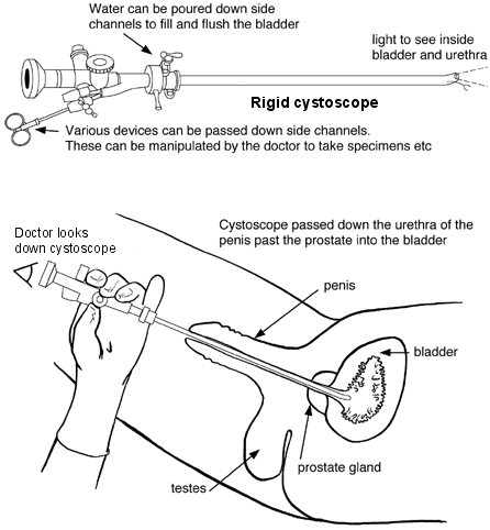
Un cistoscopio è un sottile telescopio che viene inserito nella vescica attraverso l'uretra, il tubo che porta l'urina dalla vescica all'esterno del corpo. Ci sono due tipi. Il cistoscopio può essere osservato direttamente, ma trasmette anche immagini a uno schermo del computer in modo che possano essere viste da altri membri del team. Entrambi i tipi di cistoscopio hanno canali laterali attraverso i quali possono essere inseriti vari strumenti sottili. Ad esempio, un piccolo campione (biopsia) può essere prelevato dalla parete della vescica utilizzando uno strumento che può pizzicare il tessuto e riportarlo giù lungo il canale laterale.
Tipi di cistoscopia
Un cistoscopio flessibile è il più comunemente usato. È un telescopio sottile, flessibile e a fibre ottiche che ha uno spessore simile a quello di una matita. Essendo flessibile, passa facilmente lungo le curve dell'uretra. La punta flessibile può anche essere spostata facilmente in modo che si possa vedere l'intero rivestimento della vescica.
Cistoscopio flessibile
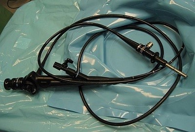
© Di Michael Reeve, tramite Wikimedia Commons
Un cistoscopio rigido è un telescopio sottile, solido e dritto. Permette a una maggiore varietà di dispositivi di passare attraverso i canali laterali, quindi può essere utilizzato per una gamma più ampia di procedure.
Diagramma di un cistoscopio rigido e come viene eseguita una cistoscopia
Procedura di cistoscopia
La cistoscopia viene solitamente eseguita come procedura ambulatoriale. Ciò significa che le persone tornano a casa lo stesso giorno e non è richiesto il pernottamento. Di solito viene eseguita mentre si è svegli. Ad alcune persone viene somministrato un sedativo per aiutarle a rilassarsi.
L'apertura dell'uretra (alla fine del pene o all'esterno della vagina) e la pelle circostante verranno pulite. Un po' di 'gel' viene quindi spruzzato nell'apertura dell'uretra. Il gel di solito contiene un anestetico locale per intorpidire il rivestimento dell'uretra. Questo aiuta il cistoscopio a passare nell'uretra con il minor disagio possibile.
Cistoscopia maschile e femminile
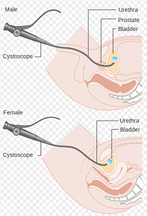
Il cistoscopio viene quindi spinto delicatamente nella vescica e il rivestimento dell'uretra e della vescica viene esaminato attentamente. L'acqua sterile viene fatta passare attraverso un canale laterale nel cistoscopio per riempire lentamente la vescica. Questo rende più facile vedere il rivestimento della vescica e la parete della vescica. Man mano che la vescica si riempie, provoca la sensazione di dover urinare, il che può essere scomodo.
Il cistoscopio viene quindi estratto delicatamente. Se è stata eseguita una biopsia, il campione verrà inviato per essere testato e osservato al microscopio. Possono passare diverse settimane prima che il referto della biopsia torni al medico.
In alcuni casi viene somministrata un'anestesia generale quando viene eseguita una cistoscopia, in particolare se viene utilizzato un cistoscopio rigido. In alcuni casi viene somministrata un'anestesia spinale che intorpidisce tutta la metà inferiore del corpo.
Quanto tempo richiede una cistoscopia?
Una cistoscopia dura circa 5-10 minuti se serve solo a guardare all'interno della vescica. Può durare più a lungo se viene eseguita una procedura - per esempio, prelevare un campione (biopsia) dal rivestimento della vescica.
La cistoscopia è dolorosa?
Le cistoscopie di solito non sono dolorose, soprattutto perché viene utilizzato un anestetico locale per inserire il cistoscopio. Possono essere scomode, specialmente quando la vescica è stata riempita d'acqua.
Complicazioni di una cistoscopia
La maggior parte delle cistoscopie viene eseguita senza alcun problema.
Per le successive 24 ore potrebbe esserci una lieve sensazione di bruciore durante la minzione e una sensazione di bisogno di urinare più spesso del solito. L'urina potrebbe anche apparire rosa a causa di un lieve sanguinamento, soprattutto se è stata effettuata una biopsia.
Occasionalmente, un infezione urinaria si sviluppa poco dopo una cistoscopia. Questo può causare un alta temperatura (febbre) e dolore durante la minzione.
Raramente, il cistoscopio può danneggiare o perforare la vescica. Dopo aver effettuato una cistoscopia, è consigliabile consultare un medico se:
Il dolore o il sanguinamento è grave.
Il dolore o il sanguinamento dura più di due giorni.
Si sviluppano sintomi di infezione, come la febbre.
Preparazione per una cistoscopia
Di solito non sono necessarie particolari preparazioni prima di una cistoscopia flessibile. Di solito è possibile mangiare e bere normalmente in anticipo. Non è necessario svuotare la vescica. Una cistoscopia rigida richiede normalmente un'anestesia generale, quindi verranno fornite indicazioni su quando smettere di mangiare e bere e cosa fare.
Dopo una cistoscopia
Dopo una cistoscopia flessibile, è normale avere bisogno di svuotare la vescica con urgenza e poi tornare a casa senza necessità di supporto aggiuntivo. Dopo una cistoscopia rigida, può essere utilizzato un catetere per svuotare la vescica e verranno fornite le normali indicazioni riguardo all'anestesia generale (attendere in ospedale per alcune ore per riprendersi, non poter guidare fino a casa, necessitare di supporto a casa per 24 ore).
Scelte del paziente per Esami delle urine e della vescica
Test e indagini
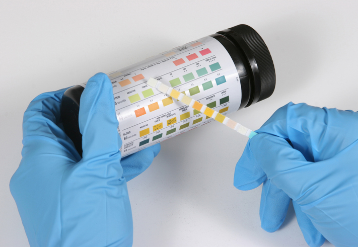
Test con striscia reattiva delle urine
Un test delle strisce reattive delle urine utilizza una striscia di carta speciale immersa in un campione di urine. Il risultato è disponibile quasi immediatamente. A volte viene chiamato test rapido delle urine.
di Dr Philippa Vincent, MRCGP

Test e indagini
Cistouretrografia minzionale
Un cistouretrogramma minzionale (MCUG/VCUG) produce immagini a raggi X della vescica e delle vie urinarie. Nota: le informazioni di seguito sono solo una guida generale. Le disposizioni e il modo in cui i test vengono eseguiti possono variare tra i diversi ospedali. Segui sempre le istruzioni fornite dal tuo medico o dall'ospedale locale.
di Dr Laurence Knott
Domande frequenti
A cosa serve un cistoscopio?
Un cistoscopio è un sottile telescopio inserito nella vescica attraverso l'uretra. Aiuta i medici a guardare all'interno della vescica e dell'uretra, e ha anche canali laterali attraverso i quali possono essere passati vari strumenti sottili per eseguire procedure come la rimozione di calcoli vescicali o l'esecuzione di biopsie.
Quanto tempo richiede una cistoscopia?
Una cistoscopia standard, se effettuata solo a scopo di esame, di solito dura circa 5-10 minuti. Tuttavia, se vengono eseguite procedure aggiuntive, come una biopsia del rivestimento della vescica o la rimozione di piccoli calcoli, la durata sarà più lunga.
Una cistoscopia richiede una preparazione speciale?
Di solito, non è necessaria una preparazione specifica per una cistoscopia flessibile; puoi mangiare e bere normalmente e non è necessario svuotare la vescica in anticipo. Se è prevista una cistoscopia rigida, che spesso comporta un'anestesia generale, verranno fornite istruzioni specifiche riguardo a cosa mangiare, bere e cosa fare.
Cosa dovrei aspettarmi dopo una cistoscopia?
Dopo una cistoscopia flessibile, probabilmente sentirai un bisogno urgente di svuotare la vescica prima di tornare a casa e di solito non avrai bisogno di ulteriore supporto. Per una cistoscopia rigida, soprattutto con anestesia generale, potrebbe essere utilizzato un catetere e riceverai consigli sul recupero dall'anestesia, come non guidare e avere bisogno di supporto a casa per 24 ore.
Quando dovrei cercare un parere medico dopo una cistoscopia?
Anche se la maggior parte delle cistoscopie non presenta problemi, dovresti consultare un medico se avverti dolore intenso o sanguinamento, o se il dolore o il sanguinamento durano più di due giorni. Dovresti anche contattare il tuo medico se sviluppi sintomi di un'infezione, come la febbre.
Ulteriori letture e riferimenti
- Zhang ZS, Wang XL, Xu CL, et al; La musica riduce il panico: uno studio iniziale sull'ascolto della musica preferita migliora il disagio e l'ansia dei pazienti maschi durante la cistoscopia flessibile. J Endourol. 2014 Giu;28(6):739-44. doi: 10.1089/end.2013.0705. Epub 2014 Mar 31.
- Gee JR, Waterman BJ, Jarrard DF, et al; Cistoscopia flessibile e rigida nelle donne. JSLS. 2009 Apr-Giu;13(2):135-8.
- Engelsgjerd JS, Deibert CM; Cistoscopia
- DeGeorge KC, Holt HR, Hodges SC; Cancro della vescica: Diagnosi e trattamento. Am Fam Physician. 15 Ott 2017;96(8):507-514.
Informazioni sull'autoreVisualizza il profilo completo

Dr Philippa Vincent, MRCGP
Medico di base, Autore medico
MB BS, Bsc, MRCGP (2000), DCH, DFSRH, DRCOG
Dr Philippa Vincent è un medico di base del NHS che lavora nel nord di Londra.
Informazioni sul recensoreVisualizza il profilo completo

Dr Colin Tidy, MRCGP
Medico di base, Autore medico
MBBS, MRCGP, MRCP (Paediatrics), DCH
Il Dr Colin Tidy è un medico del NHS, con sede nell'Oxfordshire.
Storia dell'articolo
Le informazioni su questa pagina sono scritte e revisionate da clinici qualificati.
Articolo disponibile anche in Inglese, Tedesco, Spagnolo, Francese, Italiano, Portoghese, Hindi, Ebraico, Arabo, and Svedese.
Prossima revisione prevista: 8 ago 2028
10 Ago 2023 | Ultima versione

Chiedi, condividi, connettiti.
Esplora le discussioni, fai domande e condividi esperienze su centinaia di argomenti di salute.

Non ti senti bene?
Valuta i tuoi sintomi online gratuitamente
Iscriviti alla newsletter di Patient
La tua dose settimanale di consigli sulla salute chiari e affidabili - scritti per aiutarti a sentirti informato, sicuro e in controllo.
Abbonandoti accetti i nostri Informativa sulla Privacy. Puoi annullare l'iscrizione in qualsiasi momento. Non vendiamo mai i tuoi dati.
Più in test e indagini
- Test di coagulazione del sangue
- Gruppi sanguigni e tipi
- Screening del seno
- Angiografia cerebrale
- Screening cervicale
- Campionamento dei villi coriali
- Colonscopia
- Angiografia coronarica
- Scansione DEXA
- Scansione al gallio
- Gastroscopia
- Test genetici
- Test dell'udito
- Urografia endovenosa
- Biopsia renale
- Test PSA
- Test del seme
- Spirometria
- Test con striscia reattiva delle urine
- Cistouretrografia minzionale