DEXA scan
Peer reviewed by Dr Hayley Willacy, FRCGP Last updated by Dr Colin Tidy, MRCGPLast updated 20 Mar 2023
Meets Patient’s editorial guidelines
- DownloadDownload
- Share
- Language
- Discussion
- Audio Version
- Add to preferred sources on Google
In this series:OsteoporosisBisphosphonatesCalcium-rich foodVitamin D deficiencyPreventing steroid-induced osteoporosis
DEXA scans (also called DXA scans or bone density scans) are used to check the density of bones. This test uses X-rays to show how strong bones are. A DEXA scan is different from a bone scan, which used radioactive chemicals to create a picture of the bones.
At a glance
A DEXA scan measures your bone density using low-energy X-rays.
Dense bones are stronger and less likely to break.
The scan usually takes 5 to 20 minutes while you lie on a couch.
DEXA scans use a very low level of X-ray radiation.
You do not need special preparation; avoid clothes with metal.
It may be advised if you have had a fracture or are at risk of 'thinning' bones.
DEXA scans are not advised for women who are pregnant.
Note: the information below is a general guide only. The arrangements, and the way tests are performed, may vary between different hospitals. Always follow the instructions given by your doctor or local hospital.
What is a DEXA scan?
DEXA stands for 'dual-energy X-ray absorptiometry'. DEXA (also sometimes known as DXA) is a test that measures the density of bones. Density means how much of something there is in a certain amount of space. The denser the tissue, the less X-rays pass through.
Air and water are less dense than solid things such as bone. This is because the particles which make air and water are not held closely together. In general, the more dense the bone, the stronger it is, and the less likely it is to break (fracture).
There are two different types of DEXA scanning devices:
Central DEXA devices are large machines that can measure bone density in the centre of your skeleton, such as your hip and spine.
Peripheral DEXA devices are smaller, portable machines that are used to measure bone density on the periphery of your skeleton, such as your wrist, heel or finger. These are mainly to get an idea about whether further tests are needed, as they are not as accurate as the larger DEXA machines.
How does a DEXA scan work?
A DEXA scan uses low-energy X-rays. A machine sends X-rays from two different sources through the bone being tested. Bone blocks a certain amount of the X-rays. The more dense the bone is, the less X-rays get through to the detector. By using two different X-ray sources rather than one it greatly improves the accuracy in measuring the bone density.
The amount of X-rays that comes through the bone from each of the two X-ray sources is measured by a detector. This information is sent to a computer which calculates a score of the average density of the bone. A low score indicates that the bone is less dense than it should be, some material of the bone has been lost and it is more prone to fracture.
How is a DEXA scan done?
You lie on your back on a couch and are asked to keep still while an X-ray detector (the 'scanner') comes over the area to be tested. An X-ray machine fires X-rays towards the detector. The bones commonly scanned are the bones of the back (the vertebrae), hip and wrist.
Smaller peripheral scanners are available in some places and can be used to check the bone mass density of the heel, wrist or finger.
How long does a DEXA scan take?
The scan usually takes between 5 and 20 minutes, depending on which part of your body is being examined and whether a central or peripheral scanner is being used. There is no 'tunnel' to pass through as there is in other types of scans such as an MRI or CT scan, so it should not affect people who do not like being in enclosed spaces.
DXA scanner
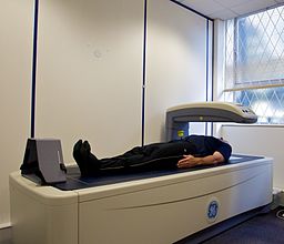
© Nick Smith photography (ALSPAC website), via Wikimedia Commons
Are DEXA scans safe?
DEXA scans use a very low level of X-ray radiation. This means it is safe for the technician doing the scan to stay in the room with you. (In standard X-ray tests, the technician has to stay behind a protective screen.)
Preparing for a DEXA scan
You do not need to do any special preparation prior to a DEXA scan. You can normally remain fully clothed, although you will need to avoid or remove clothes with metal in them (for example, zips, belts, buttons). You may also be asked to remove jewellery for the scan. In some places, you may be given a gown to wear.
Who should have a DEXA scan?
A DEXA scan may be advised if you have had a fracture of a bone after a minor injury. It may also be advised if you are considered at increased risk of 'thinning' of the bones (osteoporosis) and therefore at increased risk of having a fracture in future.
If your doctor thinks you have risk factors for osteoporosis, they may use a risk calculator such as one called FRAX® or QFracture®. This gives an idea of how likely you are to fracture your bones after a minor knock. If your risk is at a medium level, your doctor would then arrange a DEXA scan. This enables them to gain a clearer picture of your risk and then to decide whether you need any treatment.
DEXA scans are also used to monitor whether treatment for osteoporosis is working.
DEXA scans are not advised for women who are pregnant. You should also not have a DEXA scan within two weeks of certain other types of scans - for example, those using contrast dye.
For further information, see the separate leaflet called Osteoporosis.
Patient picks for Imaging

Tests and investigations
DMSA scan
A DMSA scan uses a radioactive chemical to create specialised pictures of the kidneys. It can help to show whether the kidneys are damaged or scarred. Note: the information below is a general guide only. The arrangements, and the way tests are performed, may vary between different hospitals. Always follow the instructions given by your doctor or local hospital.
by Dr Doug McKechnie, MRCGP
Tests and investigations
Ultrasound scan
An ultrasound scan is a painless test that uses sound waves to create images of organs and structures inside your body. It is a very commonly used test. As it uses sound waves it is thought to be very safe. Doppler and duplex scans are used to visualise blood or fluids flowing through the body. Note: the information below is a general guide only. The arrangements, and the way tests are performed, may vary between different hospitals. Always follow the instructions given by your doctor or local hospital.
by Dr Toni Hazell, FRCGP
Frequently asked questions
What is the purpose of a DEXA scan?
A DEXA scan is used to measure the density of bones. This measurement helps determine how strong your bones are and how likely they are to break (fracture). It can be advised if you've already had a fracture from a minor injury, or if you're considered at high risk of osteoporosis (thinning of the bones).
Does a bone density test show cancer?
The article states that a DEXA scan measures bone density to assess the risk of fractures or monitor osteoporosis treatment. It does not mention that a DEXA scan is used to detect cancer.
Do I need to do anything special to prepare for a DEXA scan?
You typically don't need any special preparation. You can usually remain fully clothed, but you will need to avoid or remove any clothing with metal, such as zips, belts, or buttons. You might also be asked to take off jewellery, and in some cases, you may be given a gown to wear.
Who should not have a DEXA scan?
DEXA scans are not recommended for pregnant women. Additionally, you should not have a DEXA scan within two weeks of having other types of scans that involve the use of contrast dye.
What is the difference between central and peripheral DEXA devices?
Central DEXA devices are large machines that measure bone density in the core parts of your skeleton, like your hip and spine. Peripheral DEXA devices are smaller, portable machines used for areas like the wrist, heel, or finger. Peripheral devices mainly indicate if further testing is needed, as they are not as accurate as the larger central machines.
Further reading and references
- Osteoporosis: assessing the risk of fragility fracture; NICE Clinical Guideline (August 2012, updated February 2017)
- Management of osteoporosis and the prevention of fragility fractures - A national clinical guideline; Scottish Intercollegiate Guidelines Network (SIGN - January 2021)
- Osteoporosis - prevention of fragility fractures; NICE CKS, July 2021 (UK access only)
- Clinical guideline for the prevention and treatment of osteoporosis; National Osteoporosis Guideline Group (updated September 2021)
About the authorView full bio

Dr Colin Tidy, MRCGP
General Practitioner, Medical Author
MBBS, MRCGP, MRCP (Paediatrics), DCH
Dr Colin Tidy is an NHS Doctor, based in Oxfordshire.
About the reviewerView full bio

Dr Hayley Willacy, FRCGP
General Practitioner, Medical Author
MBChB (1992), DRCOG, DFFP, MRCOG (Part 1) MRCGP (2007), DFSRH (2013), MSc - medical education (2020)
Dr Hayley Willacy was an NHS GP working in northwest England, who retired from clinical practice in 2022 after 30 years.
Article history
Article also available in English, German, Spanish, French, Italian, Portuguese, Hindi, Hebrew, Arabic, and Swedish.
Next review due: 18 Mar 2028
20 Mar 2023 | Latest version
Join the conversation
Share your thoughts or experiences with others in our patient communities.

Ask, share, connect.
Browse discussions, ask questions, and share experiences across hundreds of health topics.

Feeling unwell?
Assess your symptoms online for free
Sign up to the Patient newsletter
Your weekly dose of clear, trustworthy health advice - written to help you feel informed, confident and in control.
By subscribing you accept our Privacy Policy. You can unsubscribe at any time. We never sell your data.
More in tests and investigations
- Antibody and antigen tests
- Arterial blood gases
- Biopsy
- Blood groups and types
- Bone marrow biopsy and aspiration
- Coronary angiography
- Cystoscopy
- Down's syndrome screening
- Electroencephalograph
- Fine-needle aspiration
- Gastroscopy
- Hyponatraemia
- Hysteroscopy
- Intravenous urography
- Liver function tests
- Newborn screening test
- Pregnancy screening tests
- Proteinuria
- Routine kidney function blood test
- Voiding cystourethrogram