
Do I need an MRI for my back pain?
Revisione paritaria di Dr Colin Tidy, MRCGPAutore Scan.com Pubblicato originariamente 16 Jul 2026
Rispetta le linee guida editoriali
- ScaricaScarica
- Condividi
- Language
- Discussione
- Versione audio
- Aggiungi alle fonti preferite su Google
Back pain is one of the most commonly searched health topics online in the UK. Experiencing back pain can mean time off work and an inability to exercise, play sports, and take part in the activities you enjoy. Most back pain eases with time and rest, while other types require an MRI scan to get a diagnosis and the right treatment. But how do you know when you need an MRI for back pain?
Sponsorizzato
Scan.com
Understand what’s causing your lower back pain with a private MRI scan. Scan.com gives you fast, easy access to MRIs without a GP referral. Book an appointment at a clinic near you and receive your results in just a few days.

If you’ve had weeks of persistent back or spine pain coupled with vague advice to ‘rest and see’ or anxiety over what might be wrong, you’re probably wondering what happens next. Do you need a scan? If so, which one, and how do you go about getting one? Should you wait for an NHS appointment, or should you opt for a private back MRI scan?
Here’s everything you need to know.
What is an MRI scan and how does it work?
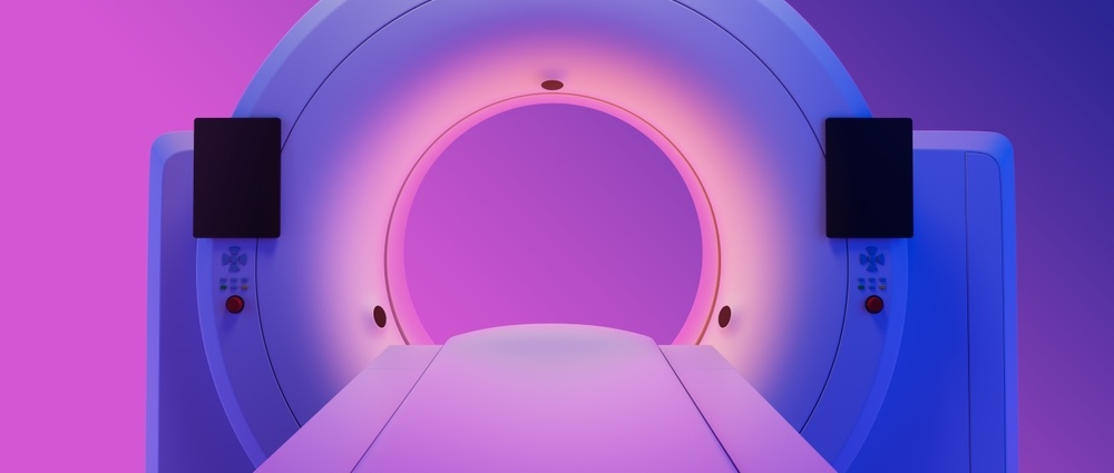
risonanza magnetica (MRI) stands for magnetic resonance imaging, and an MRI uses powerful magnetic fields and radio waves to create detailed images of the inside of the body. An MRI scanner is a large, doughnut-shaped machine, and during the scan, patients lie flat on a table that slowly moves into the scanner.
Once in the machine, the magnetic field is switched on and off, and as it does so, hydrogen atoms in the body temporarily misalign. This creates energy that computers detect and turn into an image. Different tissue types produce distinct levels of energy, and the resulting images clearly show the bones, soft tissues, organs, and fluids. Consultant radiologists can then differentiate healthy from unhealthy tissue on these images and identify areas of concern.
Unlike CTs and X–rays, MRIs don’t use radiation, and you won't feel anything as the scan takes place. However, due to the closed-in nature of the scanning machine and the loud noise from the magnets, they may be unsuitable for people with claustrophobia. MRIs take 30-90 minutes, and you’ll be required to remain still throughout.
If you struggle to remain still for any length of time, you may find an MRI difficult. Patients with magnetic metal implants, plates, and screws cannot undergo MRI scans because of the dangers posed by the magnets used in the procedure.
What can an MRI actually show in your back?
Any part of the spine can be scanned, but the most common type of back scan is a lumbar spine MRI that visualises the lower back.
A lumbar spine MRI shows the following in great detail:
Vertebrae - the bones of the spine, 33 in total, stacked on top of one another. An MRI can show fractures, bone abnormalities, and damage to the surrounding muscles, ligaments, and tendons.
Intervertebral discs - shock-absorbing discs found between each vertebra, made up of a soft, jelly-like centre and a tough exterior. MRIs show disc degeneration and herniation.
Facet joints - small joints between each vertebra that allow movement and stability. MRI scans can identify facet joint degeneration, arthritis and inflammation.
Spinal canal - the hollow canal that runs through the centre of the spine and houses the spinal cord. An MRI can detect narrowing, compression and inflammation within the spinal canal.
Spinal cord and nerves - MRIs are used to detect pinched or compressed nerves.
MRIs cannot always detect the source of pain and discomfort, and they can sometimes be inconclusive. In these cases, another scan, such as a CT, might be recommended. MRIs can also increase the risk of incidental findings, unexpected, symptomless findings that are usually harmless, but that can lead to unnecessary treatment.
MRI vs. X-rays vs. CT: which scan is right for back pain?
Doctors use different scans to diagnose the cause of back pain: MRIs, CTs, and X-rays.
Risonanza magnetica
MRI is the gold standard scan for imaging the back and spine. It creates high-quality images of soft tissues, is non-invasive, doesn’t use radiation, and is safe for most people.
MRI scans are used to diagnose conditions affecting the vertebrae, discs, spinal cord, and nerves, including fractures, spinal cord abnormalities, slipped discs, narrowing of the spinal canal, dolore neuropatico, muscular damage, infections, and cancro.
However, they can take an hour or longer and aren’t suitable if you have certain metallic implants, stents, or plates and may be difficult if you have claustrophobia or struggle to remain still for any length of time.
tomografia computerizzata
CT scans are quick and take just a few minutes, and are often used in emergencies to assess spinal damage following accidents and collisions.
They can detect minute fractures, but as they use radiation, they’re less likely to be used to diagnose non-emergency or soft tissue injuries. However, if you’re unsuitable for an MRI scan, your doctor may suggest you have a CT scan instead.
radiografia
radiografie are sometimes used as a ‘first-line’ scan, as they’re quick and useful for detecting fractures, breaks, and degeneration.
They can also show artrite, inflammation, and tumours and can be used to diagnose abnormalities in the curvature of the spine. However, they use radiation, and they’re not reliable for diagnosing problems with the discs, spinal cord, or nerves.
When it comes to CT vs MRI vs X-ray for back pain, MRI remains the gold standard if back pain needs investigating.
When do doctors recommend an MRI for back pain?
Most back pain doesn’t warrant an immediate investigation, and knowing what does and when to get a back scan can be difficult.
If you have back pain that has persisted for six weeks or more, or hasn’t responded to conservative treatments such as rest, over-the-counter pain relief, or physiotherapy, a back pain MRI scan could benefit you.
You can also benefit from an MRI if you have a condition such as sciatica, osteoporosi, or arthritis that needs monitoring or is getting worse, your doctor suspects you have a condition affecting your spine or spinal cord, you’ve previously had cancer, you’re going through cancer treatment or you have a family history of cancer.
When an MRI probably won’t change anything
If you’ve had back pain for less than six weeks and you don’t have any red flags that require urgent diagnostic testing - see below - having an MRI scan is unlikely to be of any benefit. Having an unnecessary scan may cause anxiety due to the risk of incidental findings and may lead to over-treatment.
Red flags: symptoms that mean you should act now
Certain symptoms should be investigated urgently.
Back pain red flags include:
Severe back pain that starts suddenly and has no clear cause.
Back pain following a fall, accident or collision.
Back pain that’s constant, feels worse at night in bed or doesn’t get better with rest.
Weakness, numbness or persistent tingling in the arms or legs that’s getting worse.
Weakness or numbness in the buttocks, groin or inner thighs (saddle anaesthesia).
Loss of bladder or bowel control.
An inability to urinate.
A high temperature, fever, or chills.
Perdita di peso inspiegabile.
Noticeable changes to the shape of your back or the curvature of your spine.
If you have any of these symptoms, speak to your GP or seek urgent medical attention. The sooner anything serious is discovered, the sooner treatment can start.
If you have any of these symptoms, speak to your GP or seek urgent medical attention. The sooner anything serious is discovered, the sooner treatment can start.
How to get an MRI: NHS vs private scans
If you have back pain that’s persistent and not getting better, your GP may refer you for a diagnostic MRI scan. NHS waiting times for diagnostics vary by region in the UK, but they can be long, and you may find yourself waiting several weeks or months. Waiting for a scan delays your diagnosis and treatment.
Private scans offer a fast alternative to NHS waiting lists. You can book a private MRI scan without a GP referral through Scan.com, and you’ll have your scan images and radiologist’s report within a few days.
Understanding MRI results
Once you’ve had your scan, the images are sent to a consultant radiologist for interpretation. They’ll write a report that includes a diagnosis, if appropriate, and details of any suggested further investigations.
Common terms and diagnoses associated with an MRI of the back include:
Disc bulge - protrusion of the soft, jelly-like centre of a spinal disc between each vertebra..
Disc herniation - slipped disc
Malattia degenerativa del disco - wear and tear of the shock-absorbing tissue between each vertebra.
Nerve root compression - pinched nerves.
Stenosi spinale - narrowing of the spinal canal.
Foraminal stenosis - narrowing of the bony openings (foramina) between the vertebrae where the nerves exit the spinal cord.
Infiammazione - swelling and fluid build-up.
Muscle, ligament, and tendon tears.
Fratture ossee.
It’s advised that you take your MRI images and report to your GP or specialist to discuss your next steps.
What comes next: treatment options after diagnosis
Most back pain eases with treatment, and once you have a diagnosis, knowing what treatment will benefit you the most is easier. Treatment options range from physiotherapy and pain management to interventional injections of nerve blockers. Surgical options can be offered, but they are typically used as a last resort when other options haven’t been successful.
If you have back pain that won’t go away, a scan can provide clarity and make decision-making easier.
Scelte del paziente per Test di screening

Ossa, articolazioni e muscoli
Dolore al ginocchio che non passa: quando è il momento di fare una scansione?
Il dolore al ginocchio è molto comune, ma non è sempre facile individuare cosa stia causando i tuoi sintomi. Il dolore al ginocchio può iniziare come un lieve fastidio che peggiora gradualmente, oppure improvvisamente dopo un infortunio e non scompare. Molte persone convivono con un dolore persistente al ginocchio per mesi o anni, assumendo antidolorifici o riposando la gamba quando necessario. Ma il dolore al ginocchio a lungo termine può interferire con la tua qualità di vita e la capacità di lavorare, e influenzare la tua partecipazione e il tuo divertimento in attività sportive e ricreative. Il dolore persistente al ginocchio, o il dolore che non migliora, potrebbe richiedere ulteriori indagini. Fare una scansione fornirà chiarezza su cosa stia causando i tuoi sintomi e quali opzioni di trattamento potrebbero essere disponibili per te. Ma a volte è difficile sapere quando fare una scansione e quale tipo scegliere. Scan.com aiuta a rendere il tutto più semplice.
di Scan.com
Test e indagini
La guida completa per ottenere una scansione
X-rays, CTs, MRIs, and ultrasounds are all types of medical scans. Millions of patients receive medical scans each year in the UK, but unless you’ve had one, the process can feel confusing. How do you get referred? Which scan do you need? Which body parts can be scanned and what can they diagnose?
di Scan.com
Ulteriori letture e riferimenti
- NHS Back pain
- NICE CKS Sciatica - Red flags, June 2026 (UK access only)
- MRI and low back pain; MedlinePlus
Informazioni sull'autoreVisualizza il profilo completo

Scan.com
Informazioni sul recensoreVisualizza il profilo completo

Dr Colin Tidy, MRCGP
Medico di base, Autore medico
MBBS, MRCGP, MRCP (Paediatrics), DCH
Il Dr Colin Tidy è un medico del NHS, con sede nell'Oxfordshire.
Storia dell'articolo
Le informazioni su questa pagina sono revisionate da clinici qualificati.
Articolo disponibile anche in Inglese, Tedesco, Spagnolo, Francese, Italiano, Portoghese, Hindi, Ebraico, Arabo, and Svedese.
Next review due: 16 Jul 2029
16 Jul 2026 | Pubblicato originariamente
Autore:
Scan.comRevisione paritaria di
Dr Colin Tidy, MRCGP

Chiedi, condividi, connettiti.
Esplora le discussioni, fai domande e condividi esperienze su centinaia di argomenti di salute.

Non ti senti bene?
Valuta i tuoi sintomi online gratuitamente
Iscriviti alla newsletter di Patient
La tua dose settimanale di consigli sulla salute chiari e affidabili - scritti per aiutarti a sentirti informato, sicuro e in controllo.
Abbonandoti accetti i nostri Informativa sulla Privacy. Puoi annullare l'iscrizione in qualsiasi momento. Non vendiamo mai i tuoi dati.
Più in test e indagini
- I test allergici privati valgono la pena?
- Un esame del sangue potrebbe rilevare il cancro ovarico due anni prima
- Un nuovo esame del sangue può rilevare precocemente il cancro ai polmoni?
- Le donne lesbiche e bisessuali hanno ancora bisogno di fare il Pap test?
- Le scansioni cerebrali precoci possono prevedere la prognosi della sclerosi multipla
- Tutto quello che devi sapere sullo screening del cancro
- Come alleviare l'ansia durante l'attesa dei risultati degli esami medici
- Come superare la paura degli esami del sangue
- Come superare la paura del pap test
- È facile fare un test HIV?
- Un nuovo test del sangue potrebbe diagnosticare l'insufficienza cardiaca in modo più precoce
- Prepararsi per il pap test come sopravvissuto alla violenza sessuale
- Opzioni di test PSA
- Test delle urine per il cancro alla prostata potrebbe rivelare chi necessita di trattamento urgente
- Cosa potrebbe significare un risultato anomalo del test di Pap
- Che cos'è un esame del sangue CA125?
- Che cos'è il test del sangue per gli anticorpi COVID-19 e dovrei farlo?
- Qual è la differenza tra allergia e intolleranza?
- Cosa aspettarsi durante uno screening mammario
- Perché le persone transgender hanno difficoltà ad accedere allo screening cervicale?