Cisti pilari
Epidermoid and sebaceous cysts
Revisione paritaria di Dr Toni Hazell, FRCGPUltimo aggiornamento di Dr Philippa Vincent, MRCGPUltimo aggiornamento 20 Nov 2021
Rispetta le linee guida editoriali
- ScaricaScarica
- Condividi
- Language
- Discussione
- Versione audio
- Aggiungi alle fonti preferite su Google
Professionisti Medici
Gli articoli di riferimento professionale sono progettati per essere utilizzati dai professionisti della salute. Sono scritti da medici del Regno Unito e basati su prove di ricerca, linee guida del Regno Unito ed europee. Potresti trovare il Cisti epidermoidali e pilari articolo più utile, o uno dei nostri altri articoli sulla salute.
What are epidermoid and pilar cysts?
Epidermoid or pilar cysts are very common and are usually of no clinical significance.
Epidermoid cysts (also known as epithelial, epidermal or sebaceous cysts) are intradermal or subcutaneous tumours. Epidermoid cysts may occur anywhere on the body but occur most often on the face, scalp, neck, back and scrotum. Although they are most commonly known as sebaceous cysts, they actually do not involve the sebaceous gland.1
Pilar cysts (also called trichilemmal cysts) are clinically indistinguishable from epidermal cysts. They contain keratinous material, are often multiple and there may be an autosomal dominant inheritance.2
Who gets epidermoid and pilar cysts? (Epidemiology)2
Pilar cysts and epidermoid cysts are most common types of skin cyst.
They are usually sporadic but can occur as part of an autosomal dominant trait.
They are more common in women than men.
They are very slow growing.
Epidermoid cysts are also usually sporadic but can occur as part of an autosomal dominant trait.
They tend to occur between the ages of 20 and 40 and are twice as common in men than women.
Babies often develop tiny epidermoid cysts, which spontaneously resolve, called milia, colloquially known as "baby acne" or "milk spots" (though neither of these names is accurate).
Up to 1% of epidermoid cysts have been shown to transform to squamous cell carcinoma or basal cell carcinoma.
Immunosuppressants such as imiquimod and cyclosporine have been shown to cause epidermoid cysts.
Epidermoid cyst on the neck
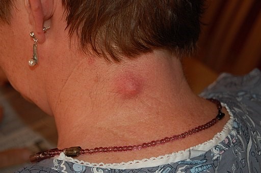
© Steven Fruitsmaak (Own work) - close-up view, CC BY-SA 3.0, via Wikimedia Commons
Epidermoid and pilar cyst symptoms
Most often they present as a painless skin lump.
They may present with discharge of an offensive thick creamy material.
If they become infected, they are red, inflamed and painful.
Lesions of the genitals can be painful during intercourse and cause problems with walking or wearing underwear. They can also interfere with micturition.
Epidermoid and pilar cysts appear as firm, round, mobile, flesh-coloured to yellow or white subcutaneous nodules of variable size.
A central pore or punctum may tether the cyst to the overlying epidermis and a thick cheesy material can sometimes be expressed.
In people with dark skin, the cysts may also be pigmented.
Sites affected by epidermoid and pilar cysts
The sites most commonly affected are, in descending order of frequency, the face, the trunk, the neck, the extremities and the scalp.
Cysts of the genitals are less common and may appear as a mass in the vulva, the clitoris, the penis, the scrotum, or the perineum. In cultures which practise female circumcision (or female genital mutilation) cysts on the vulva are common.
The ocular and oral mucosae can also be affected and cysts have been reported on the palpebral conjunctivae, on the lips, on the buccal mucosa, on and under the tongue and even on the uvula. These are not sites with hair follicles.
Cysts may occur on the extremities. Subungual cysts can cause changes in the nails, such as onycholysis and subungual hyperkeratosis, which may be mistaken for psoriasis or onychomycosis. These cysts also produce changes in the nails (such as pincer nails) erythema, oedema, tenderness and pain.
Palmoplantar lesions represent a unique subset of epidermoid cysts.
Diagnosi differenziale
Other causes of skin cysts.
A lipoma tends to be larger and is very soft.
A neurofibroma is hard and may be multiple.
An abscess is hot and red and may resemble an infected sebaceous cyst.
A Merkel's cell carcinoma may look like a pilar or epidermoid cyst but occurs in older people and tends to be very fast growing.
Multiple cysts in a teenager may suggest Gardner's syndrome.
Diagnosing epidermoid and pilar cysts (investigations)
Usually the diagnosis is clear and no investigations are required. If malignancy is suspected, excision and histology are required.
Malattie associate
Epidermoid cysts are a feature of Gardner's syndrome which is an autosomal dominant condition comprising familial polyposis coli, cutaneous cysts and osteomas or other soft-tissue tumours. The cysts tend to occur at an earlier age than usual (presenting most often in the early teenage years). The face may be involved but the extremities tend to be affected more than the trunk.
Epidermoid and pilar cyst treatment
Most people with an epidermoid or pilar cyst do not need medical attention.
If a cyst is uncomplicated then no treatment is usually advised. The cyst may disappear spontaneously, leaving no trace. Even the most skilful excision will leave a permanent scar.
An infected cyst will usually require an oral antibiotic effective against staphylococci - for example, flucloxacillin. The infection may be mixed and, in lesions of the scalp and anogenital area, anaerobic flora are more likely.
If the cyst has ruptured, the contents can be expressed. However, the cyst may well re-form.
If the cyst is troublesome or if the patient, after counselling, is eager to have it removed then the entire cyst should be excised as a surgical procedure. However, this is usually not available on the NHS unless it is causing significant symptoms.
Excision of an epidermoid or pilar cyst
Vedi anche l'articolo separato Chirurgia minore in cure primarie.
The usual preliminaries of informed consent are assumed.
Information includes the warning that the cyst may recur if all the wall is not removed.
Appropriate aseptic precautions should be taken.
Local anaesthetic such as 1% lidocaine with adrenaline (epinephrine) is used (note contra-indications to use of adrenaline (epinephrine) in certain areas). The area is infiltrated with local anaesthetic, being careful not to puncture the cyst.
Make a careful and superficial incision over the cyst avoiding rupture of the cyst. Care should be taken to remove all the cyst wall if the cyst is ruptured.
Toothed forceps can be used to grip the skin - a blunt dissection of adhesions allows mobilisation of the cyst and removal. A Volkmann spoon or scissors are useful for the blunt dissection.
If the cyst is large and leaves a significant defect then a resorbable subcutaneous suture should be used to close a potential space for haematoma to form.
The diagnosis of an epidermoid or pilar cyst is usually obvious. However, excised tissue should always be submitted for histology.
Complications with epidermoid or pilar cysts
Dislike of the cosmetic appearance
Infezione.
Malignant change is rare (causes rapid growth, friability and bleeding).
Prognosi
They will usually grow slowly and only need removal if causing symptoms. They tend to recur if incised rather than excised.
Aggiornamenti esclusivi per i professionisti sanitari
Rimani informato con gli ultimi aggiornamenti clinici, approfondimenti professionali e linee guida basate su evidenze. La newsletter Patient Pro seleziona contenuti essenziali per i professionisti sanitari—consegnati direttamente nella tua casella di posta.
Abbonandoti accetti i nostri Informativa sulla Privacy. Puoi annullare l'iscrizione in qualsiasi momento. Non vendiamo mai i tuoi dati.
Ulteriori letture e riferimenti
- Al Aboud DM, Yarrarapu SNS, Patel BC; Pilar Cyst. StatPearls, August 2021.
- Trichilemmal cyst; DermNet NZ.
- Pilar cyst; Società di Dermatologia delle Cure Primarie.
- Zito PM, Scharf R; Epidermoid Cyst.
- Al Aboud DM, Yarrarapu SNS, Patel BC; Pilar Cyst.
Informazioni sull'autoreVisualizza il profilo completo

Dr Philippa Vincent, MRCGP
Medico di base, Autore medico
MB BS, Bsc, MRCGP (2000), DCH, DFSRH, DRCOG
Dr Philippa Vincent è un medico di base del NHS che lavora nel nord di Londra.
Informazioni sul recensoreVisualizza il profilo completo

Dr Toni Hazell, FRCGP
MBBS, BSc, FRCGP, DFSRH, Dip GU med, DRCOG, DCH (London, UK, 2000)
La Dott.ssa Toni Hazell si è laureata presso la St. Mary’s Hospital Medical School e ha completato il suo VTS al Northwick Park Hospital.
Storia dell'articolo
Le informazioni su questa pagina sono scritte e revisionate da clinici qualificati.
Articolo disponibile anche in Inglese, Tedesco, Spagnolo, Francese, Italiano, Portoghese, Hindi, Ebraico, Arabo, and Svedese.
Prossima revisione prevista: 19 Nov 2026
20 Nov 2021 | Ultima versione

Chiedi, condividi, connettiti.
Esplora le discussioni, fai domande e condividi esperienze su centinaia di argomenti di salute.

Non ti senti bene?
Valuta i tuoi sintomi online gratuitamente
Altro in dermatologia
- Albinismo
- Farmaci antifungini
- Dermatite atopica ed eczema
- Campbell de Morgan spot
- Cellulite ed erisipela
- Sindrome di Chediak-Higashi
- Infezione da Coxsackievirus
- Eczema su mani e piedi
- Eritema anulare centrifugo
- Erythema marginatum rheumatica
- Herpes genitale in gravidanza
- HIV e disturbi della pelle
- Iperidrosi
- Lichen planus
- Micetoma
- Fotodermatosi
- Lesioni pigmentate della pelle
- Noduli cutanei e sottocutanei
- Smallpox