ERCP
Endoscopic retrograde cholangiopancreatography
Peer reviewed by Dr Hayley Willacy, FRCGP Last updated by Dr Colin Tidy, MRCGPLast updated 13 Nov 2023
Meets Patient’s editorial guidelines
- DownloadDownload
- Share
- Language
- Discussion
- Audio Version
- Add to preferred sources on Google
In this series:GallstonesCholecystitisAcute pancreatitis
ERCP is a procedure that uses an endoscope and X-rays to look at the bile duct and the pancreatic duct. ERCP can also be used to remove gallstones or take small samples of tissue for analysis (a biopsy).
Note: the information below is a general guide only. The arrangements and the way tests are performed may vary between different hospitals. Always follow the instructions given by the doctor or local hospital.
At a glance
An ERCP is a procedure that uses an endoscope and X-rays to diagnose and treat conditions.
It can diagnose and treat problems like gallstones and inflammation of the pancreas.
A thin, flexible tube is passed through the mouth to look inside and perform treatments.
A sedative is usually given to make you drowsy and relaxed during the procedure.
You must not eat for six hours before the ERCP.
After the procedure, avoid driving, operating machinery, or drinking alcohol for 24 hours.
Contact a doctor immediately if you have worsening tummy pain, fever, difficulty breathing, or vomit blood afterwards.
What is an ERCP?
ERCP stands for 'endoscopic retrograde cholangiopancreatography'. ERCP is a very useful procedure, as it can be used both to diagnose and to treat various conditions, such as:
Acute pancreatitis - inflammation of the pancreas that develops quickly over a few days.
Chronic pancreatitis - inflammation of the pancreas that is more persistent.
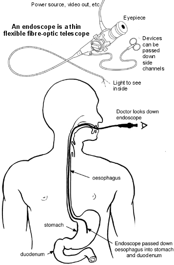
An endoscope is a thin, flexible tube. It is passed through the mouth, into the gullet (oesophagus) and down towards the stomach and the first part of the gut after the stomach (duodenum).
The endoscope contains fibre-optic channels which allow light to shine down so the doctor can see inside. Cholangiopancreatography means X-ray pictures of the bile duct and pancreatic duct. These ducts do not show up very well on ordinary X-ray pictures. However, if a contrast dye that blocks X-rays is injected into these ducts then X-ray pictures will show up these ducts clearly.
Some dye is injected through an opening called 'the papilla' back up into the bile and pancreatic ducts (a 'retrograde' injection). This is done via a plastic tube in a side channel of the endoscope. X-ray pictures are then taken.
Prior to having an ERCP, blood pressure, heart rate, respiration and oxygen levels will be recorded. If a person has diabetes, the blood sugar level will also be checked and recorded. Blood tests may also be needed prior to the ERCP. The procedure will be discussed in detail and a consent form signed once the procedure is fully understood.
What happens during an ERCP procedure?
ERCP
The doctor may numb the back of the throat by spraying on some local anaesthetic, or may give a lozenge to suck. A sedative will usually be given by an injection into a vein in the back of the hand or arm. The sedative causes drowsiness and relaxes but it is not like a general anaesthetic.
While lying on n a couch, lying on one side, the first section of the endoscope is swallowed. Modern endoscopes are quite thin (thinner than an index finger) and quite easy to swallow. The doctor then gently pushes it down the oesophagus into the stomach and duodenum.
The doctor looks down the endoscope via an eyepiece or on a TV monitor which is connected to the endoscope. Air is passed down a channel in the endoscope into the stomach and the first part of the duodenum. This enables the lining to be seen more easily. It often causes a feeling of 'fullness' and wanting to belch.
The endoscope also has a 'side channel' down which various tubes or instruments can pass. These can be manipulated by the doctor who can do various things. For example:
Inject a dye into the bile and pancreatic ducts. X-ray pictures taken immediately after the injection of dye show up the detail of the ducts. This may show narrowing (stricture), stuck gallstones, tumours pressing on the ducts, etc.
Take a small sample (biopsy) from the lining of the duodenum, stomach, or pancreatic or bile duct near to the opening called the papilla. The biopsy sample can be looked at under the microscope to check for abnormal tissue and cells.
If the X-rays show a gallstone stuck in the duct, the doctor can widen the opening of the papilla to let the stone out into the duodenum. A stone can be grabbed by a 'basket' or left to be passed out with the stools (faeces).
If the X-rays show a narrowing or blockage in the bile duct, the doctor can put a stent inside to open it wide. A stent is a small wire-mesh or plastic tube. This then allows bile to drain into the duodenum in the normal way. The stent can remain permanently in place without being aware that it is there.
The endoscope is gently pulled out when the procedure is finished. An ERCP can take anything from 30 minutes to over an hour, depending on what is done.
Preparing for an ERCP
Instructions will be provided by the hospital department before an ERCP. The sort of instructions given include:
Not eating for six hours before the procedure. (Small sips of water may be allowed up to two hours before the procedure.)
Advice about any usual medication that should be stopped before the procedure.
Taking antibiotics before the procedure. This depends on the reason for having this test done.
What to expect after an ERCP
If the procedure was done just to obtain X-ray pictures then most people are ready to go home after resting for a few hours. Driving, operating machinery or drinking alcohol must be avoided for 24 hours after having the sedative.
If going home on the same day as the procedure, it is important to have somebody to accompany home and to stay with for 24 hours until the effects of the sedative have fully worn off.
Most people are able to resume normal activities after 24 hours. Because of the effect of the sedative, most people remember very little about the procedure. A short hospital stay may be needed following a procedure such as removing a gallstone or inserting a small wire-mesh or plastic tube (a stent).
Complications of an ERCP
Most ERCPs are done without any problems. Some people have a mild sore throat for a day or so afterwards. Feeling tired or sleepy for several hours, caused by the sedative, is common.
Uncommon complications include the following:
Occasionally, the endoscope causes some damage to the gut, bile duct or pancreatic duct. This may cause bleeding, infection and, rarely, perforation. If any of the following occur within 48 hours after an ERCP, a doctor should be contacted immediately:
Tummy (abdominal) pain - in particular, if it becomes gradually worse and is different or more intense to any 'usual' indigestion pains or heartburn.
Raised temperature (fever).
Difficulty breathing.
Bringing up (vomiting) blood.
Inflammation of the pancreas (pancreatitis) sometimes occurs after ERCP. This can be serious in some cases.
The risk of complications is higher if already in poor general health. The benefit from this procedure needs to be weighed up against the small risk of complications.
It may still be possible to perform ERCP during pregnancy, providing certain precautions are taken. Alternatively, it may be possible to delay it or use another type of procedure.
Patient picks for Endoscopy

Surgery and procedures
Nasoendoscopy
A nasoendoscopy is a test to look inside the nose (nasal passage), the back of the throat (pharynx) and the voice box (larynx). It's sometimes called a flexible nasal endoscopy or FNE. Note: the information below is a general guide only. The arrangements and the way tests are performed may vary between different hospitals. Always follow the instructions given by your doctor or local hospital.
by Dr Doug McKechnie, MRCGP

Surgery and procedures
Hysteroscopy
Hysteroscopy is a procedure which uses a thin tube-like telescope to see inside the womb (uterus). It can also allow doctors to do some minor operations to the uterus.
by Dr Philippa Vincent, MRCGP
Frequently asked questions
What is the primary difference between acute and chronic pancreatitis?
Acute pancreatitis refers to inflammation of the pancreas that develops quickly over a few days. In contrast, chronic pancreatitis describes an inflammation of the pancreas that is more persistent and long-lasting.
How do doctors visualise the bile and pancreatic ducts during an ERCP if they don't show up well on regular X-rays?
Doctors inject a special contrast dye into these ducts. This dye blocks X-rays, making the bile and pancreatic ducts clearly visible on the X-ray pictures taken during the procedure.
What should I do if I have diabetes and need an ERCP?
If you have diabetes, your blood sugar level will be checked and recorded before the ERCP procedure. It's important to discuss your diabetes management with the healthcare team ahead of time.
Will I be fully unconscious during an ERCP?
No, you will usually be given a sedative, which causes drowsiness and helps you relax. However, it is not the same as a general anaesthetic, meaning you won't be fully unconscious.
How long does a stent stay in place after being inserted during an ERCP?
A stent inserted during an ERCP is designed to remain permanently in place. Most people are not aware that it is there once it has been fitted.
Why might I need to take antibiotics before an ERCP?
Taking antibiotics before an ERCP depends on the specific reason you are having the test done. Your hospital department will provide advice on whether this is necessary for your situation.
What are some of the immediate symptoms I should look out for after an ERCP that would require contacting a doctor?
You should contact a doctor immediately if you experience abdominal pain that gradually worsens, especially if it's different or more intense than usual indigestion, a raised temperature (fever), difficulty breathing, or vomiting blood within 48 hours after your ERCP.
Further reading and references
- Riff BP, Chandrasekhara V; The Role of Endoscopic Retrograde Cholangiopancreatography in Management of Pancreatic Diseases. Gastroenterol Clin North Am. 2016 Mar;45(1):45-65. doi: 10.1016/j.gtc.2015.10.009.
- Meseeha M, Attia M; Endoscopic Retrograde Cholangiopancreatography. StatPearls Publishing, August 2023.
- Ribeiro IB, do Monte Junior ES, Miranda Neto AA, et al; Pancreatitis after endoscopic retrograde cholangiopancreatography: A narrative review. World J Gastroenterol. 2021 May 28;27(20):2495-2506. doi: 10.3748/wjg.v27.i20.2495.
- Azab M, Bharadwaj S, Jayaraj M, et al; Safety of endoscopic retrograde cholangiopancreatography (ERCP) in pregnancy: A systematic review and meta-analysis. Saudi J Gastroenterol. 2019 Nov-Dec;25(6):341-354. doi: 10.4103/sjg.SJG_92_19.
About the authorView full bio
Updated by

Dr Colin Tidy, MRCGP
General Practitioner, Medical Author
MBBS, MRCGP, MRCP (Paediatrics), DCH
Dr Colin Tidy is an NHS Doctor, based in Oxfordshire.
About the reviewerView full bio

Dr Hayley Willacy, FRCGP
General Practitioner, Medical Author
MBChB (1992), DRCOG, DFFP, MRCOG (Part 1) MRCGP (2007), DFSRH (2013), MSc - medical education (2020)
Dr Hayley Willacy was an NHS GP working in northwest England, who retired from clinical practice in 2022 after 30 years.
Join the conversation
Share your thoughts or experiences with others in our patient communities.

Ask, share, connect.
Browse discussions, ask questions, and share experiences across hundreds of health topics.

Feeling unwell?
Assess your symptoms online for free
Sign up to the Patient newsletter
Your weekly dose of clear, trustworthy health advice - written to help you feel informed, confident and in control.
By subscribing you accept our Privacy Policy. You can unsubscribe at any time. We never sell your data.