Nevo sebaceo
Revisione paritaria di Dr Philippa Vincent, MRCGPUltimo aggiornamento di Dr Doug McKechnie, MRCGPUltimo aggiornamento 24 maggio 2023
Rispetta le linee guida editoriali
- ScaricaScarica
- Condividi
- Language
- Discussione
- Versione audio
- Aggiungi alle fonti preferite su Google
Professionisti Medici
Gli articoli di riferimento professionale sono progettati per essere utilizzati dai professionisti della salute. Sono scritti da medici del Regno Unito e basati su prove di ricerca, linee guida del Regno Unito ed europee. Potresti trovare uno dei nostri articoli sulla salute più utile.
Synonyms: naevus sebaceous, naevus sebaceous of Jadassohn, verrucous epidermal naevus, organoid naevus. Note the American spelling 'nevus'.
What is a sebaceous naevus?
A sebaceous naevus is a congenital skin lesion which usually manifests as a bald patch on the scalp of the baby. The area of hair loss looks orange or yellowish, with a velvety feel and appearance. It is usually oval or circular. In adolescence, under the influence of sex hormones, it becomes bumpy and warty, with an unpleasant verrucoid appearance.
Throughout teenage years and early adulthood they usually stay quiescent. Sometimes they enlarge in later adulthood and, rarely, undergo neoplastic change which on the whole involves benign tumours.
Sebaceous naevi are hamartomas, meaning they arise from tissues normally found at that site but they grow in a disorganised way. They have a common embryological origin of the pilosebaceous-apocrine unit and therefore contain sebaceous glands, hair follicles and sweat glands. Hence the term sebaceous naevus is a little inaccurate: some pathologists prefer the term verrucous epidermal naevus or organoid naevus.1 Although they contain elements of hair follicles, normal terminal hair follicles are absent making the area look hairless. Most are solitary, with the scalp being the most common site.
Scalp sebaceous naevus
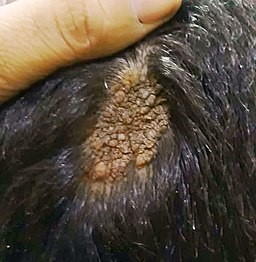
© Mohammad2018, CC BY-SA 4.0, via Wikimedia Commons
Sebaceous naevus (Epidemiology)
Estimated prevalence is 0.3% of all newborns worldwide, with an equal sex distribution.2
Sebaceous naevus symptoms (presentation)
Usually, a single hairless patch (round or linear) is noted on the scalp at birth, or shortly thereafter.
The classical velvety tan or yellow-orange appearance may be noted.
The lesion may appear more prominent in newborns, due to the effects of maternal sex hormones.
In adolescents the lesion tends to take on its nodular verrucoid appearance due to the influence of sex hormones.
In later life there may be overgrowth of components of the lesion indicating possible benign or malignant tumours developing within the lesion.
Diagnosi differenziale
The appearance of sebaceous naevi is fairly characteristic but the following lesions may have a similar appearance or affect similar areas of the body:
Aplasia cutis congenita.
Naevus syringocystadenomatosus papilliferous.
Juvenile xanthogranulomata.
Solitary mastocytoma.
Altre cause di alopecia.
Indagini
Biopsy may be considered for diagnostic purposes or to investigate the nature of tumours that arise within them.
Malattie associate
About a third of people with a sebaceous naevus have systemic abnormalities affecting the brain, eye or skeletal systems. When these occur it is termed sebaceous naevus syndrome.3 In these cases the naevi follow the lines of Blaschko; the more extensive the cutaneous involvement, the greater the risk of extracutaneous manifestations.4 Subsequent problems can include infantile epilepsy, developmental delay and intellectual disability.
Sebaceous naevus treatment and management5
Due to the difficulty of quantifying the risk of malignant transformation, some specialists advocate full-thickness skin excision before adolescence.6 Others recommend watchful waiting.5
Some cases will in any case not be suitable for excision due to their size and location. These should be subjected to regular review.
Photodynamic therapy can play a role in removing the lesion.7
Seek the advice of a dermatologist and/or plastic surgeon to help decide on the need for, and timing of, excision.
Any lesion that changes its appearance significantly, ulcerates, becomes painful, bleeds or shows other signs of potential malignancy, should be referred to a dermatologist.
Complicazioni
Poor cosmetic appearance.
Development of a benign or malignant tumour within the lesion.
Complications associated with removal of the lesion.
Prognosi
Most lesions remain quiescent throughout adult life, the only concern being cosmetic. Two case series studying nearly 1,300 sebaceous naevi have found that 15% undergo neoplastic change.8 9
Of these 15%, 80% develop foci of benign neoplasms such as trichoblastomas and syringocystadenomas. The remaining 20% develop malignant changes, with basal cell carcinomas making up half of those, squamous cell carcinomas a quarter.
Usually, any malignant change occurs in adulthood but it has been reported in children as young as 8 years.6 Malignancy can be diagnosed in the same way as other malignant skin tumours, with dermoscopy playing its usual role.10
Aggiornamenti esclusivi per i professionisti sanitari
Rimani informato con gli ultimi aggiornamenti clinici, approfondimenti professionali e linee guida basate su evidenze. La newsletter Patient Pro seleziona contenuti essenziali per i professionisti sanitari—consegnati direttamente nella tua casella di posta.
Abbonandoti accetti i nostri Informativa sulla Privacy. Puoi annullare l'iscrizione in qualsiasi momento. Non vendiamo mai i tuoi dati.
Ulteriori letture e riferimenti
- Simi CM, Rajalakshmi T, Correa M; Clinicopathologic analysis of 21 cases of nevus sebaceus: a retrospective study. Indian J Dermatol Venereol Leprol. 2008 Nov-Dec;74(6):625-7.
- Moody MN, Landau JM, Goldberg LH; Nevus sebaceous revisited. Pediatr Dermatol. 2012 Jan-Feb;29(1):15-23. doi: 10.1111/j.1525-1470.2011.01562.x. Epub 2011 Oct 13.
- Laura FS; Epidermal nevus syndrome. Handb Clin Neurol. 2013;111:349-68. doi: 10.1016/B978-0-444-52891-9.00041-5.
- Asch S, Sugarman JL; Epidermal nevus syndromes. Handb Clin Neurol. 2015;132:291-316. doi: 10.1016/B978-0-444-62702-5.00022-6.
- Nevo sebaceo; DermNet NZ
- Rosen H, Schmidt B, Lam HP, et al; Management of nevus sebaceous and the risk of Basal cell carcinoma: an 18-year review. Pediatr Dermatol. 2009 Nov-Dec;26(6):676-81. doi: 10.1111/j.1525-1470.2009.00939.x. Epub 2009 Jul 20.
- Babilas P, Szeimies RM; The use of photodynamic therapy in dermatology. G Ital Dermatol Venereol. 2010 Oct;145(5):613-30.
- Idriss MH, Elston DM; Secondary neoplasms associated with nevus sebaceus of Jadassohn: a study of 707 cases. J Am Acad Dermatol. 2014 Feb;70(2):332-7. doi: 10.1016/j.jaad.2013.10.004. Epub 2013 Nov 20.
- Cribier B, Scrivener Y, Grosshans E; Tumors arising in nevus sebaceus: A study of 596 cases. J Am Acad Dermatol. 2000 Feb;42(2 Pt 1):263-8.
- Enei ML, Paschoal FM, Valdes G, et al; Basal cell carcinoma appearing in a facial nevus sebaceous of Jadassohn: dermoscopic features. An Bras Dermatol. 2012 Jul-Aug;87(4):640-2.
Informazioni sull'autoreVisualizza il profilo completo

Dr Doug McKechnie, MRCGP
Scrittore Medico
MA, MBBS, MSc, DRCOG, MRCP(UK), MRCGP(2021), FHEA
Il dottor Doug McKechnie è un medico di base del NHS che lavora a Londra. Lavora a tempo pieno in ambito clinico ed è anche Vice Responsabile del modulo di Pratica Clinica e Professionale presso la Scuola di Medicina dell'University College London.
Informazioni sul recensoreVisualizza il profilo completo

Dr Philippa Vincent, MRCGP
Medico di base, Autore medico
MB BS, Bsc, MRCGP (2000), DCH, DFSRH, DRCOG
Dr Philippa Vincent è un medico di base del NHS che lavora nel nord di Londra.
Storia dell'articolo
Le informazioni su questa pagina sono scritte e revisionate da clinici qualificati.
Articolo disponibile anche in Inglese, Tedesco, Spagnolo, Francese, Italiano, Portoghese, Hindi, Ebraico, Arabo, and Svedese.
Prossima revisione prevista: 12 maggio 2028
24 maggio 2023 | Ultima versione

Chiedi, condividi, connettiti.
Esplora le discussioni, fai domande e condividi esperienze su centinaia di argomenti di salute.

Non ti senti bene?
Valuta i tuoi sintomi online gratuitamente
Altro in dermatologia
- Cimici dei letti
- Naevus blu
- Dermatite artefattiva
- Nevo epidermico e le sue sindromi
- Erythema marginatum rheumatica
- Psoriasi guttata
- Nevo di Halo
- Cicatrici cheloidi
- Filariasi linfatica
- Melanoma maligno della pelle
- Nocardia
- Fotodermatosi
- Lesioni pigmentate della pelle
- Malattia ungueale psoriasica
- Pioderma gangrenoso
- Angioma stellato
- Sindrome della pelle ustionata da stafilococco
- Vene varicose