Ecocardiogramma
Revisione paritaria di Dr Colin Tidy, MRCGPUltimo aggiornamento di Dr Toni Hazell, MRCGPUltimo aggiornamento 22 Nov 2022
Rispetta le linee guida editoriali
- ScaricaScarica
- Condividi
- Language
- Discussione
- Versione audio
- Aggiungi alle fonti preferite su Google
An echocardiogram is an ecografia of the heart. It is sometimes just called an 'echo'. Ultrasound is a very high-frequency sound that you cannot hear but it can be emitted and detected by special machines. The scan can give accurate pictures of the heart muscle, the heart chambers and structures within the heart such as the valves.
A colpo d'occhio
An echocardiogram uses ultrasound to create moving pictures of your heart.
It can show how well your heart is working and how blood flows through it.
The test is painless, takes 15-30 minutes, and requires no special preparation.
You will need to undress to the waist and lie on a couch.
A sonographer will place a probe with lubricating jelly on your chest.
Results are sent to the doctor who requested the test.
Do not contact your GP for results of a test requested by a consultant.
In questo articolo:
Scelte video per Esami cardiaci
Continua a leggere sotto
What does an echocardiogram show?
An echocardiogram can be carried out for many different reasons. It may be done to check how well your heart is working after major heart problems such as a heart attack, or to look at how well the valves are moving inside the heart. An echocardiogram can also help to see any fluid that may have collected around the heart and may be used to see if symptoms such as shortness of breath are caused by a cardiac cause such as heart failure.
How accurate is a echocardiogram?
Interpreting an echocardiogram is a skilled job. The sonographer will interpret the images and write a report, which will be sent to the requesting clinician (GP or consultant). In some cases a consultant may want to carry out an echocardiogram themselves, to get a first-hand look at the images.
How is an echocardiogram done?
Torna ai contenutiThe test is painless and takes about 15-30 minutes. You may have to turn on to your side during the test so that the operator can scan the heart from different angles.
You will need to undress to the waist and lie on the couch. A probe is placed on your chest (it is a bit like a very thick blunt pen). Also, a substance called a contrast agent (lubricating jelly) is put on your chest so the probe makes good contact with the skin. The probe is connected by a wire to the ultrasound machine and monitor. Pulses of ultrasound are sent from the probe through the skin towards your heart. The ultrasound waves then 'bounce back' (echo) from the heart and various structures in the heart.
Echocardiogram procedure
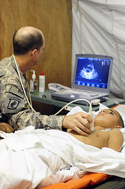
© Tech Sgt Luke Thelen, via Wikimedia Commons
The echoes are detected by the probe and are sent to the echocardiogram machine. They are displayed as a picture on the monitor. The picture is constantly updated so the scan can show movement as well as structure. (For example, the valves of a heart opening and closing.) The operator moves the probe around over the skin surface to obtain views from different angles. Some abnormalities can be seen quite clearly. For example, damaged heart valves, thickened heart muscle, some congenital heart defects, etc.
You do not need any special preparation before the test. You eat and drink normally before and after the test. Continue to take your usual medication.
Continua a leggere sotto
Doppler echocardiography
Torna ai contenutiThis type of echocardiogram can measure variations in blood flow through your heart. For example, if can detect any abnormal blood flows next to a damaged valve. It can assess how well the heart valves are working. You do not need any special preparation before this test.
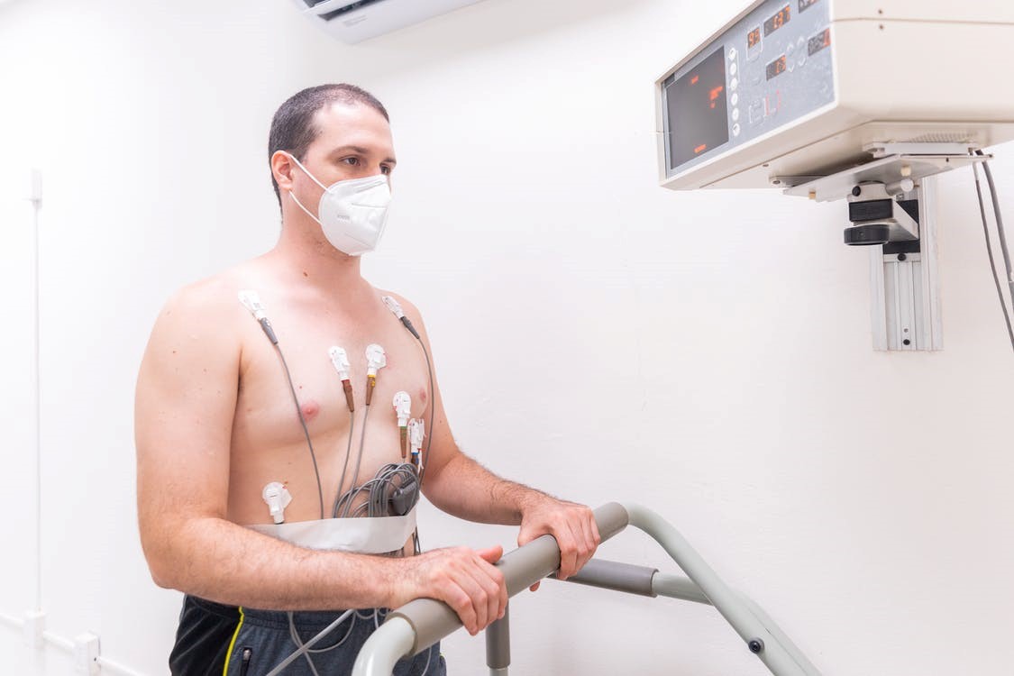
Stress echocardiogram
Torna ai contenutiThis test is done to show how well your heart responds to 'stress' such as exercise. In this test your doctor may do an echocardiogram, as described above, during or soon after exercise. Or you may be given a medication that causes the heart to beat harder and faster.
Continua a leggere sotto
Transoesophageal echocardiogram
Torna ai contenutiIn this test you swallow a probe that is attached to a thin tube connecting it to an ultrasound machine. This views the heart from within the gullet (oesophagus) which lies just behind the heart. This can give a clearer view of the heart than normal echocardiography. It is done in situations where a very detailed picture is needed. For example, to assess valves before surgery is done to repair damaged valves, or to assess the extent of infection of a heart valve.
How long does it take to get echocardiogram results?
Torna ai contenutiThis varies locally. Results will go back to the requesting clinician and it is their responsibility to give them to the patient. So, if your echocardiogram was requested by a consultant, do not ring your GP for the result. Wait for your next consultant appointment, where you will get the result, or ring your consultant's secretary if you have a query. Hospital consultants should not ask patients to go to their GP for the results of tests that the consultant has requested.
Scelte del paziente per Esami cardiaci

Test e indagini
Elettrocardiogramma ambulatoriale
L'elettrocardiogramma ambulatoriale monitora il tuo cuore mentre svolgi le tue normali attività. Aiuta a rilevare frequenze e ritmi cardiaci anomali (aritmie). Le disposizioni e il modo in cui vengono eseguiti i test possono variare tra i diversi ospedali. Segui sempre le istruzioni fornite dal tuo medico o dall'ospedale locale.
di Dr Toni Hazell, MRCGP
Test e indagini
Exercise Tolerance Testing
An exercise tolerance test (ETT) records the electrical activity of the heart whilst exercising. It is most useful in patients who experience chest pain on exertion. It is also used to detect whether heart rhythm abnormalities can be brought on by exercise.
di Dr Philippa Vincent, MRCGP
Domande frequenti
What is the gel used during an echocardiogram for?
A lubricating jelly, also called a contrast agent, is put on your chest during an echocardiogram. This helps the probe make good contact with your skin, allowing the ultrasound waves to pass through effectively.
Will I feel anything when the ultrasound waves go through my heart?
No, the test is painless. Pulses of ultrasound are sent from the probe through your skin towards your heart, and these waves 'bounce back' to create an image, but you will not feel them passing through.
Why would a Transoesophageal echocardiogram be chosen over a regular echocardiogram?
A Transoesophageal echocardiogram offers a clearer and more detailed view of the heart than a standard echocardiogram because the probe is swallowed and positions the ultrasound close to the heart, from within the gullet. This method is used when a very detailed picture is essential, for example, to assess heart valves before surgery or to check for infection.
What kind of problems can an echocardiogram identify?
An echocardiogram can identify a variety of problems, including damaged heart valves, thickened heart muscle, and some congenital heart defects. It can also show how well your heart is working after a heart attack, check valve movement, and detect fluid around the heart.
Who interprets the echocardiogram images?
A skilled sonographer interprets the images from the echocardiogram and compiles a report. This report is then sent to the clinician who requested the test, such as your GP or a consultant. In some cases, a consultant might perform the echocardiogram themselves.
Do I need to do anything to prepare for an echocardiogram?
No, you do not need any special preparation before an echocardiogram. You can eat and drink normally, and continue to take your usual medication as prescribed.
Ulteriori letture e riferimenti
- Sengupta PP, Khandheria BK; Transoesophageal echocardiography. Heart. 2005 Apr;91(4):541-7.
- Mohamed AA, Arifi AA, Omran A; The basics of echocardiography. J Saudi Heart Assoc. 2010 Apr;22(2):71-6. doi: 10.1016/j.jsha.2010.02.011. Epub 2010 Mar 1.
Continua a leggere sotto
Informazioni sull'autoreVisualizza il profilo completo

Dr Toni Hazell, MRCGP
MBBS, BSc, MRCGP, DFSRH, Dip GU med, DRCOG, DCH (London, UK, 2000)
La Dott.ssa Toni Hazell si è laureata presso la St. Mary’s Hospital Medical School e ha completato il suo VTS al Northwick Park Hospital.
Informazioni sul recensoreVisualizza il profilo completo

Dr Colin Tidy, MRCGP
Medico di base, Autore medico
MBBS, MRCGP, MRCP (Paediatrics), DCH
Il Dr Colin Tidy è un medico del NHS, con sede nell'Oxfordshire.
Storia dell'articolo
Le informazioni su questa pagina sono scritte e revisionate da clinici qualificati.
Prossima revisione prevista: 21 Nov 2027
22 Nov 2022 | Ultima versione

Chiedi, condividi, connettiti.
Esplora le discussioni, fai domande e condividi esperienze su centinaia di argomenti di salute.

Non ti senti bene?
Valuta i tuoi sintomi online gratuitamente
Iscriviti alla newsletter di Patient
La tua dose settimanale di consigli sulla salute chiari e affidabili - scritti per aiutarti a sentirti informato, sicuro e in controllo.
Abbonandoti accetti i nostri Informativa sulla Privacy. Puoi annullare l'iscrizione in qualsiasi momento. Non vendiamo mai i tuoi dati.