Megalencephalic leukoencephalopathy with subcortical cysts
Ultimo aggiornamento 18 Ago 2020
Rispetta le linee guida editoriali
- ScaricaScarica
- Condividi
- Language
- Discussione
- Ask Patient
- Versione audio
- Aggiungi alle fonti preferite su Google
Megalencephalic leukoencephalopathy with subcortical cysts is a progressive condition that affects brain development and function. Individuals with this condition typically have an enlarged brain (megalencephaly) that is evident at birth or within the first year of life. Megalencephaly leads to an increase in the size of the head (macrocephaly). Affected people also have leukoencephalopathy, an abnormality of the brain's white matter. White matter consists of nerve fibers covered by a fatty substance called myelin. Myelin insulates nerve cells (neurons) and promotes the rapid transmission of nerve impulses. In megalencephalic leukoencephalopathy with subcortical cysts, the myelin is swollen and contains numerous fluid-filled pockets (vacuoles). Over time, the swelling decreases and the myelin begins to waste away (atrophy). Individuals affected with this condition may develop cysts in the brain; because these cysts form below an area of the brain called the cerebral cortex, they are called subcortical cysts. These cysts can grow in size and number.
Scelte video per Condizioni geneticheVideo library
Descrizione
Megalencephalic leukoencephalopathy with subcortical cysts is a progressive condition that affects brain development and function. Individuals with this condition typically have an enlarged brain (megalencephaly) that is evident at birth or within the first year of life. Megalencephaly leads to an increase in the size of the head (macrocephaly). Affected people also have leukoencephalopathy, an abnormality of the brain's white matter. White matter consists of nerve fibers covered by a fatty substance called myelin. Myelin insulates nerve cells (neurons) and promotes the rapid transmission of nerve impulses. In megalencephalic leukoencephalopathy with subcortical cysts, the myelin is swollen and contains numerous fluid-filled pockets (vacuoles). Over time, the swelling decreases and the myelin begins to waste away (atrophy). Individuals affected with this condition may develop cysts in the brain; because these cysts form below an area of the brain called the cerebral cortex, they are called subcortical cysts. These cysts can grow in size and number.
The brain abnormalities in people with megalencephalic leukoencephalopathy with subcortical cysts affect the use of muscles and lead to movement problems. Affected individuals typically experience muscle stiffness (spasticity) and difficulty coordinating movements (ataxia). Walking ability varies greatly among those affected. Some people lose the ability to walk early in life and need wheelchair assistance, while others are able to walk unassisted well into adulthood. Minor head trauma can further impair movements and may lead to coma. Affected individuals may also develop uncontrolled muscle tensing (dystonia), involuntary writhing movements of the limbs (athetosis), difficulty swallowing (dysphagia), and impaired speech (dysarthria). More than half of all people with this condition have recurrent seizures (epilepsy). Despite the widespread brain abnormalities, people with this condition typically have only mild to moderate intellectual disability.
There are three types of megalencephalic leukoencephalopathy with subcortical cysts, which are distinguished by their signs and symptoms and genetic cause. Types 1 and 2A have different genetic causes but are nearly identical in signs and symptoms. Types 2A and 2B have the same genetic cause but the signs and symptoms of type 2B often begin to improve after one year. After improvement, individuals with type 2B usually have macrocephaly and may have intellectual disability.
Modelli di ereditarietà
The source lists the following possible inheritance patterns: Autosomal recessive; Autosomal dominant. More than one pattern can apply in different cases.
Related genes
Altri nomi
Infantile leukoencephalopathy and megalencephaly
Leukoencephalopathy with swelling and a discrepantly mild course
Leukoencephalopathy with swelling and cysts
LVM
MLC
Vacuolating leukoencephalopathy
Vacuolating megalencephalic leukoencephalopathy with subcortical cysts
Van der Knaap disease
Related patient information and medicines

Patient information leaflet
Gonfiori e protuberanze al collo
Gonfiori e protuberanze al collo are common and they have many possible causes. They may arise from the skin or from structures underneath. Key points Noduli al collo have many causes including swollen lymph nodes or salivary glands, cysts or abscesses, and tumours. Most lumps are nothing to worry about and are often linked to infections such as colds,.
Patient information leaflet
Disturbi della ghiandola pituitaria
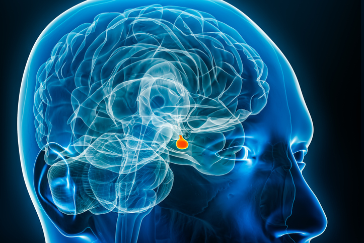
This leaflet gives a brief overview of the problema della ghiandola pituitaria and the hormones it makes. The pituitary gland is found at the base of the brain and is 'pea-sized'. The pituitary gland is an overall controller of a number of other glands in the body, overseeing the function of these organs through hormones. Some of the pituitary gland disorders.

Patient information leaflet
Mediastinoscopia
Mediastinoscopy is a type of 'keyhole' surgery that allows doctors to look inside the chest. It can be used to take samples of body tissue for further testing. Note : the information below is a general guide only. The arrangements, and the way tests are performed, may vary between different hospitals. Always follow the instructions given by your.

Patient information leaflet
Decongestionanti
Dr Krishna Vakharia , 21st February 2024 A note on pseudoephedrine-containing products. Following a review by MHRA - the safety information of all pseudoephedrine-containing medicines will be updated to provide clearer descriptions of these risks and potential risk factors for these conditions for both patients and healthcare professionals. PRES-.

Patient information leaflet
Ecografia endoscopica
Un endoscopic ultrasound scan uses an endoscope with an l'ecografia probe attached to create detailed pictures of internal organs and structures. Note : the information below is a general guida only. The arrangements and the way tests are performed, may vary between different hospitals. Always follow the instructions given by your doctor or local.

Patient information leaflet
Cisti epidermoidali e pilari
Epidermoid e pilar cysts are commonly referred to as 'sebaceous cysts' (pronounced 'seb-ay-shuss'). They are overgrowths of skin cells (called keratin) held together in a little capsule, or sac. They are harmless smooth lumps just under the surface of the skin. They are not cancerous and do not require removal unless they are bothering you by.
Ulteriori letture e riferimenti
- Megalencephalic leukoencephalopathy with subcortical cysts; MedlinePlus, National Library of Medicine.
- Arnedo T, Aiello C, Jeworutzki E, Dentici ML, Uziel G, Simonati A, Pusch M, Bertini E, Estevez R. Expanding the spectrum of megalencephalic leukoencephalopathy with subcortical cysts in two patients with GLIALCAM mutations. Neurogenetics. 2014 Mar;15(1):41-8. doi: 10.1007/s10048-013-0381-x. Epub 2013 Nov 8.
- Arnedo T, Lopez-Hernandez T, Jeworutzki E, Capdevila-Nortes X, Sirisi S, Pusch M, Estevez R. Functional analyses of mutations in HEPACAM causing megalencephalic leukoencephalopathy. Hum Mutat. 2014 Oct;35(10):1175-8. doi: 10.1002/humu.22622. Epub 2014 Aug 18.
- Ilja Boor PK, de Groot K, Mejaski-Bosnjak V, Brenner C, van der Knaap MS, Scheper GC, Pronk JC. Megalencephalic leukoencephalopathy with subcortical cysts: an update and extended mutation analysis of MLC1. Hum Mutat. 2006 Jun;27(6):505-12. doi: 10.1002/humu.20332.
- Lopez-Hernandez T, Sirisi S, Capdevila-Nortes X, Montolio M, Fernandez-Duenas V, Scheper GC, van der Knaap MS, Casquero P, Ciruela F, Ferrer I, Nunes V, Estevez R. Molecular mechanisms of MLC1 and GLIALCAM mutations in megalencephalic leukoencephalopathy with subcortical cysts. Hum Mol Genet. 2011 Aug 15;20(16):3266-77. doi: 10.1093/hmg/ddr238. Epub 2011 May 30.
- Montagna G, Teijido O, Eymard-Pierre E, Muraki K, Cohen B, Loizzo A, Grosso P, Tedeschi G, Palacin M, Boespflug-Tanguy O, Bertini E, Santorelli FM, Estevez R. Vacuolating megalencephalic leukoencephalopathy with subcortical cysts: functional studies of novel variants in MLC1. Hum Mutat. 2006 Mar;27(3):292. doi: 10.1002/humu.9407.
- Pascual-Castroviejo I, van der Knaap MS, Pronk JC, Garcia-Segura JM, Gutierrez-Molina M, Pascual-Pascual SI. Vacuolating megalencephalic leukoencephalopathy: 24 year follow-up of two siblings. Neurologia. 2005 Jan-Feb;20(1):33-40.
- Singhal BS, Gorospe JR, Naidu S. Megalencephalic leukoencephalopathy with subcortical cysts. J Child Neurol. 2003 Sep;18(9):646-52. doi: 10.1177/08830738030180091201.
Condividi i tuoi pensieri o esperienze con gli altri nel nostro comunità di pazienti.
Accesso semplice a trattamenti affidabili e servizi sanitari online.
Common conditions
Get help with common conditions through the NHS, without a GP appointment.
 Sinusite acuta
 Tonsillite batterica
 Impetigine
 Punture di insetti
 Fuoco di Sant'AntonioInfezioni delle vie urinarie (IVU)
Iscriviti alla newsletter di Patient
La tua dose settimanale di consigli sulla salute chiari e affidabili - scritti per aiutarti a sentirti informato, sicuro e in controllo.
Abbonandoti accetti i nostri Informativa sulla Privacy. Puoi annullare l'iscrizione in qualsiasi momento. Non vendiamo mai i tuoi dati.