Skin cancer types
Peer reviewed by Dr Colin Tidy, MRCGPLast updated by Dr Toni Hazell, FRCGPLast updated 24 Feb 2023
Meets Patient’s editorial guidelines
- DownloadDownload
- Share
- Language
- Discussion
- Audio Version
- Add to preferred sources on Google
This leaflet gives a general overview about skin cancer and the different skin cancer types - melanoma and non-melanoma.
At a glance
Skin cancer occurs when skin cells grow abnormally and multiply out of control.
The main types are melanoma, basal cell carcinoma (BCC), and squamous cell carcinoma (SCC).
BCC and SCC are more common, often appearing as lumps, red scaly patches, or ulcers.
Melanoma is less common but more serious and can spread to other body areas.
Most skin cancers are caused by damage from ultraviolet (UV) radiation from the sun.
Early detection leads to a better chance of successful treatment for all types.
Protect your skin from the sun, especially children, to reduce your risk.
What is cancer?
Cancer is a disease of the cells in the body. There are many different types of cell in the body, and there are many different types of cancer which arise from different types of cell. What all types of cancer have in common is that the cancer cells are abnormal and multiply out of control. See the separate leaflet called Cancer for more details.
What are the different skin cancer types?
There are different types of skin cell in the body and different types of cancer develop from them. Skin cancers are divided into:
Melanoma (malignant melanoma). This type of skin cancer develops from melanocytes. There are around 13,000 new cases in the UK each year.
Non-melanoma. These are more common than melanomas. The different types of non-melanomatous skin cancer are:
Basal cell carcinoma (BCC) - skin cancer which develops from basal cells. There are around 75,000 new cases in the UK each year.
Squamous cell carcinoma (SCC) - skin cancer which develops from keratinocytes. There are around 25,000 new cases in the UK each year.
Other - other types of skin cancer, which are rare.
A malignant tumour is a growth of tissue made up from cancer cells which continue to multiply. Malignant tumours can invade into nearby tissues and cause damage. Most types of malignant tumour also tend to spread (metastasise) to other parts of the body. Melanoma and SCC can spread (metastasise) to distant parts of the body, but a BCC can only spread locally. However, this local spread can be very damaging, particularly if the BCC is close to an important structure such as the eye.
Basal cell carcinoma (BCC)
Basal cell carcinoma - nodular type
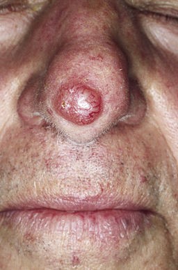
© M. Sand, D. Sand, C. Thrandorf, V. Paech, P. Altmeyer, F. G. Bechara, CC BY 2.0, via Wikimedia Commons
Basal cell skin cancer is the most common type of skin cancer in white and fair-skinned people. BCC is rare in dark-skinned people. BCC is more common in older people. Those aged over 75 years are about five times more likely to have a BCC than those people aged between 50-55 years.
A BCC typically develops on a sun-exposed area of the skin such as the head and neck. However, one can develop on any area of skin. The first sign is often a small red, pink or pearly lump which appears on previously normal skin. The lump is often dome-shaped. However, BCCs can vary in shape and colour. They usually grow very slowly and it can take many months for one to grow to a centimetre or more.
In time, the lump on the skin may crust over, ulcerate or bleed from time to time. A skin ulcer caused by a BCC is sometimes called a rodent ulcer which often looks like a small inflamed crater with a raised edge. BCCs very rarely spread (metastasise) to other parts of the body. However, untreated they will continue to grow locally and can cause damage to nearby structures. For example, a BCC on the face may erode and damage the nose or an ear.
Squamous cell carcinoma (SCC)
Squamous cell carcinoma - forearm
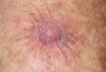
© Dermanonymous(Own work), via Wikimedia Commons
Squamous cell skin cancer is the second most common skin cancer in white and fair-skinned people. SCC is rare in dark-skinned people. Like BCCs, SCCs are more common in older people. Those aged over 75 years are about thirty-five times more likely to have an SCC than those aged between 50-55 years.
An SCC typically develops on the face - most commonly on or around the ears or lips. However any area of skin can be affected. It typically starts as a red scaly patch. It may grow into a lump which may look like a wart. An SCC may ulcerate or bleed from time to time. However, an early SCC can vary in shape, appearance and colour.
As an SCC grows larger and deeper, it damages nearby structures. For example, if left untreated, an SCC next to a nose or ear can grow into, erode and then completely destroy the nose or ear. An SCC may also spread to other areas of the body. However, this is uncommon in the early stages and most are treated before any spread occurs.
Melanoma (malignant melanoma)
Melanoma is the least common form of skin cancer, but it is the most serious. There are about 9,000 new cases of melanoma each year in the UK. Melanoma is the second most common cancer in people aged 15-34 years. It is more than twice as common in young women as it is in young men.
A typical melanoma starts as a small dark patch on the skin (similar to a mole). It can develop from a normal part of skin, or from an existing mole. A melanoma is often different to a mole in one or more of the following ways (summed up as ABCDE) - that is:
Asymmetry - the shape of a melanoma is often uneven and asymmetrical, unlike a mole which is usually round and even.
Border - the border or edges of a melanoma are often ragged, notched or blurred. A mole has a smooth well-defined edge.
Colour - the colour (pigmentation) of a melanoma is often not uniform. So there may be 2-3 shades of brown or black. A mole usually has one uniform colour.
Diameter - the size of a melanoma is usually larger than a normal mole and it continues to grow.
Evolving - any change in size, shape, colour, or elevation or any new symptom such as bleeding, itching or crusting may be due to a melanoma.
Read more about melanoma skin cancer. The section includes diagrams of melanomas and compares then to normal moles.
Bowen's disease
Bowen's disease is a very early type of skin cancer, which usually presents with a red scaly patch on the skin, usually on the lower legs. It is also known as an SCC in situ. If left alone it may develop into an SCC - this only happens to about 3-5% of those with Bowen's disease.
What causes skin cancer?
The cause of most skin cancers is sun damage to the skin. About 9 in 10 non-melanoma skin cancers and about 6 in 10 melanomas are thought to be caused by excessive exposure to the sun. In particular, past episodes of sunburn significantly increase the risk.
It is the ultraviolet (UV) radiation in the sunshine which does the damage. Skin cells which are damaged are at greater risk of becoming abnormal and cancerous.
See the leaflets called Melanoma Skin Cancer, Non-melanoma Skin Cancer and Preventing Skin Cancer, for other possible risk factors and causes.
Although skin cancer is rare in children, the amount of sun exposure during childhood is thought to increase the risk of developing skin cancers in adult life. Therefore, it is vital to protect children from too much sunshine (see below).
Understanding the skin
The skin has two layers - the epidermis and the dermis. Beneath the dermis is a layer of fat and then the deeper structures such as muscles and tendons. There are different cells in each layer of the skin.
Skin diagram cross-section view
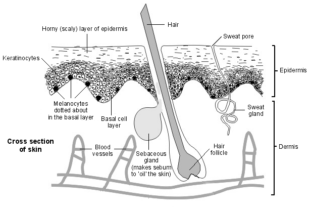
The epidermis has three main types of cell:
Basal cells. These are the bottom layer of cells in the epidermis.
Keratinocytes. These cells are in layers above the basal layer. They make a substance called keratin which is a hard wax-like material. Keratinocytes are constantly dividing and a certain number are dying at any given time. The top horny layer of the epidermis is made of dead keratinocytes which contain keratin. The top of the skin is constantly being shed and replaced by new dead cells which contain keratin.
Melanocytes. These cells are dotted about at the bottom of the epidermis. They make a pigment called melanin when skin is exposed to sun. The melanin is passed to the nearby skin cells to protect them from the sun's rays. Melanin causes the skin to tan in fair-skinned people. Dark-skinned people have more active melanocytes.
What is the treatment and prognosis for skin cancer?
All three main skin cancer types - basal cell carcinoma (BCC), squamous cell carcinoma (SCC) and melanoma - are curable if they are detected at an early stage. A small operation or other method to remove the affected area of skin is all that is required in early cases.
For non-melanoma skin cancers
The overall cure rate is over 95%, even for ones which have been present for a while. However, the larger they grow, the more difficult they are to treat. More extensive surgery or other localised treatments may be needed if they grow large or deep before they are treated.
For melanomas
The risk of spread to other parts of the body is high. This is why urgent early treatment is needed. An operation is required to remove the melanoma. If it has already spread to other areas of the body then there is less chance of a cure. Treatments such as chemotherapy, radiotherapy or immunotherapy may be used for melanomas which have spread.
How can I check for the early signs of skin cancer?
Try to get to know the site and look of the normal moles or marks on your body so that you will know if there has been any change. If you notice any new growth or new change on your skin and you do not know what it is then see a doctor. In particular, if you notice any change in the size, shape or colour of an existing mole, or if a new dark area of skin develops.
See our skin cancer checklist for more information on how to spot the signs.
How can I prevent skin cancer from developing?
Most skin cancers are caused by excessive exposure to the sun. We should all limit our sun exposure in the summer months (or all year when in hot countries nearer the equator) by:
Staying indoors or seeking the shade as much as possible between 11 am and 3 pm.
Covering up with clothes and a wide-brimmed hat when you are out in the sunshine.
Applying sunscreen of at least sun protection factor (SPF) 15 (SPF 30 for children or people with pale skin) which also has high ultraviolet A (UVA) protection.
In particular, children should be protected from the sun. Sunburn or excessive exposure to the sun in childhood is thought to be the biggest risk factor for the developing of skin cancer as an adult.
Patient picks for Skin cancer
Cancer
Non-melanoma skin cancer
Non-melanoma skin cancers are any skin cancers that are not melanoma. The term is used because melanoma is very different to other skin cancers so it's important to think of them as separate types of skin cancers. Basal cell carcinomas (BCCs) and squamous cell carcinomas (SCCs) are the main types of non-melanoma skin cancers.
by Dr Philippa Vincent, MRCGP
Cancer
Preventing skin cancer
Some skin cancers can be partly prevented by protecting yourself from the sun, not smoking, and generally taking care of your health. Some kinds of skin cancer can't be prevented however, because they are caused by something going wrong in your genes: this is beyond your control.
by Dr Oliver Starr, MRCGP
Frequently asked questions
Can skin cancer affect dark-skinned individuals?
While the article mentions that Basal Cell Carcinoma (BCC) and Squamous Cell Carcinoma (SCC) are rare in dark-skinned people, it does not state that they are impossible. The article focuses on fair-skinned individuals as the primary demographic for these types, but it does not exclude others. Melanoma can also affect all skin types.
What specifically is a 'malignant tumour'?
A malignant tumour is a growth of tissue made up of cancer cells that continue to multiply. These tumours can invade and damage nearby tissues and, in most cases, have the ability to spread (metastasise) to other parts of the body. Different types of skin cancer have varying tendencies to spread; for example, melanoma and SCC can spread distantly, while BCC typically spreads only locally.
What is Bowen's disease and how does it relate to skin cancer?
Bowen's disease is considered a very early type of skin cancer, also referred to as SCC in situ. It typically appears as a red, scaly patch on the skin, often on the lower legs. If left untreated, there is a small chance (about 3-5%) that it could develop into a more serious Squamous Cell Carcinoma (SCC).
Are there other risk factors for skin cancer besides sun exposure?
The article states that sun damage is the cause of most skin cancers, specifically mentioning that about 9 in 10 non-melanoma skin cancers and 6 in 10 melanomas are linked to excessive sun exposure, particularly past episodes of sunburn. It also mentions that more information on other possible risk factors and causes can be found in separate leaflets, implying additional factors exist even if not detailed here.
How quickly do different types of skin cancer grow?
Basal Cell Carcinoma (BCC) usually grows very slowly; it can take many months for a BCC to grow to a centimetre or more. The article does not specify growth rates for Squamous Cell Carcinoma (SCC) or Melanoma, but notes that SCC can grow larger and deeper, and melanoma involves 'evolving' changes indicating growth.
Further reading and references
- DermNet NZ
- UV forecast; Met Office
- DermIS - Dermatology Information System
- Melanoma: assessment and management; NICE Guidance (July 2015 - last updated July 2022)
- Cutaneous Melanoma: Clinical Practice Guidelines; European Society for Medical Oncology (2019)
- Basal cell carcinoma; British Association of Dermatologists, 2022
- Squamous Cell Carcinoma; British Association of Dermatologists, 2022
About the authorView full bio

Dr Toni Hazell, FRCGP
MBBS, BSc, FRCGP, DFSRH, Dip GU med, DRCOG, DCH (London, UK, 2000)
Dr. Toni Hazell qualified from St. Mary’s Hospital Medical School and did her VTS at Northwick Park Hospital.
About the reviewerView full bio

Dr Colin Tidy, MRCGP
General Practitioner, Medical Author
MBBS, MRCGP, MRCP (Paediatrics), DCH
Dr Colin Tidy is an NHS Doctor, based in Oxfordshire.
Article history
Article also available in English, German, Spanish, French, Italian, Portuguese, Hindi, Hebrew, Arabic, and Swedish.
Next review due: 7 Feb 2028
24 Feb 2023 | Latest version
Join the conversation
Share your thoughts or experiences with others in our patient communities.

Ask, share, connect.
Browse discussions, ask questions, and share experiences across hundreds of health topics.

Feeling unwell?
Assess your symptoms online for free
Sign up to the Patient newsletter
Your weekly dose of clear, trustworthy health advice - written to help you feel informed, confident and in control.
By subscribing you accept our Privacy Policy. You can unsubscribe at any time. We never sell your data.
More in cancer
- Acute myeloid leukaemia
- Bone marrow biopsy and aspiration
- Bowel cancer screening
- Cancer symptoms
- Causes of cancer
- Cervical screening
- Childhood leukaemias
- Diagnosing cancer
- End of life care
- Faecal immunochemical test
- Hodgkin's lymphoma
- Kidney cancer
- Mammogram
- Myeloma
- Preventing skin cancer
- Primary liver cancer
- Prostate cancer
- Rhabdomyosarcoma
- Testicular cancer
- Thyroid cancer